

Disclaimer: This page contains content specific to Florida Stem Cell Law, which permits specific licensed physicians to administer stem cell therapies not yet approved by the U.S. Food and Drug Administration. This content applies to providers licensed in Florida under Chapter 458 (Medical Doctors) and Chapter 459 (Osteopathic Physicians) acting within the scope of their practice.
Diagnostic ultrasound, also known as musculoskeletal sonography, is a non-ionizing imaging modality that uses high-frequency sound waves to visualize soft tissues, joints, tendons, ligaments, and fluid collections in real time. In contrast to MRI or CT scans, ultrasound offers instantaneous, dynamic visualization of structures without exposure to ionizing radiation.
Diagnostic ultrasound is integral to the clinical assessment and formulation of treatment strategies for musculoskeletal disorders. Its ability to provide real-time visualization of tissue structures in motion facilitates the identification of inflammation, tendon degeneration, joint effusion, partial tears, and structural irregularities that may contribute to chronic pain. Unlike reliance on static imaging reports, ultrasound permits immediate structural evaluation at the point of care, enabling findings to be directly correlated with a patient’s symptoms during the same consultation.
Portable ultrasound provides real-time imaging that facilitates diagnosis and treatment, thereby enhancing clinicians’ confidence and assurance in regenerative and musculoskeletal care. Cutting-edge portable systems are revolutionizing healthcare delivery, spanning from vascular health assessment to the guidance of precise interventions, thus cultivating trust in clinical decision-making.
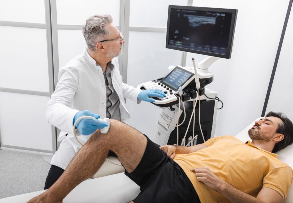
Chronic musculoskeletal pain typically arises from soft tissue degeneration rather than complete structural rupture. Ultrasound facilitates real-time visualization of tendon thickening, fiber irregularity, synovial inflammation, joint effusion, and regions of scar tissue formation, which may not be readily discernible on standard radiographs.
Because imaging is performed during the consultation, findings can be immediately correlated with the patient’s specific pain pattern. For example, shoulder motion can be assessed dynamically to evaluate rotator cuff function and impingement mechanics. Knee evaluation may reveal joint fluid accumulation or tendon degeneration that contributes to instability. This immediate structural insight helps determine whether conservative care, shockwave therapy, Platelet-Rich Plasma (PRP), exosome therapy, or stem cell therapy is most appropriate for the individual patient.
Portable ultrasound enables healthcare professionals to move beyond superficial assessments by providing detailed imaging of vascular structures. For instance, carotid artery intima-media thickness (IMT) measurements are extensively utilized in anti-aging treatments and cardiovascular risk assessment. Moreover, ultrasound guidance enhances the safety of administering regenerative therapies, including platelet-rich plasma (PRP) injections, stem cell treatments, and filler safety mapping.
In the realm of musculoskeletal care, ultrasound technology facilitates real-time visualization of tendons, ligaments, joints, and soft tissues. This facilitates clinicians in evaluating injuries, monitoring healing progress, and guiding procedures such as joint injections, tendon repairs, or regenerative therapies to enhance mobility and alleviate pain.
In addition to its diagnostic utility, ultrasound substantially improves procedural accuracy during regenerative treatments. When administering therapies such as PRP, MSC-derived exosomes, or stem cell therapy, real-time ultrasound guidance facilitates direct visualization of needle placement within the targeted tissue structures.
This enhances both safety and precision by enabling the clinician to avoid adjacent nerves and vascular structures while ensuring that biological material is accurately delivered to areas of degeneration or inflammation. Instead of relying solely on surface anatomy landmarks, ultrasound guidance verifies correct placement within tendon insertions, joint spaces, or ligament attachments. Image-guided injection techniques are widely recognized for improving procedural reliability, treatment consistency, and patient outcomes.
Diagnostic ultrasound is used to assess orthopedic, pain, and wound-healing issues. The selection of an imaging modality is contingent upon clinical examination findings, symptom duration, and treatment planning requirements.
Diagnostic ultrasound is conducted in a clinical setting utilizing a handheld transducer positioned over the area of concern. A conductive gel is applied to the skin to facilitate the transmission of sound waves through the tissue. The procedure is entirely painless and generally completed within a few minutes. Since imaging results are available immediately, structural findings can be reviewed and discussed during the same consultation, allowing direct discussion of findings and treatment alternatives without delay. Unlike MRI or CT scans, ultrasound can be safely repeated as often as necessary without any radiation exposure.
Magnetic Resonance Imaging (MRI) continues to be regarded as the gold standard for the comprehensive assessment of deep intra-articular structures. Nonetheless, diagnostic ultrasound offers several notable advantages in musculoskeletal healthcare. It enables real-time assessment of dynamic movements, provides immediate bedside imaging during clinical consultations, and offers precise guidance for regenerative injection procedures. For superficial tendons, ligaments, and soft-tissue structures, ultrasound often provides exceptional tissue resolution while remaining cost-effective and readily accessible. It is particularly well-suited for evaluating chronic soft tissue conditions and guiding regenerative interventions with high accuracy.
Not every orthopedic condition requires ultrasound evaluation. The decision to use ultrasound imaging is contingent on clinical examination findings, symptom presentation, and treatment-planning considerations. Diagnostic ultrasound is not a substitute for MRI in all cases — especially for deep intra-articular pathologies, where MRI offers superior resolution. A thorough clinical consultation is essential to ascertain whether ultrasound imaging is the most appropriate modality for your particular condition.